Bronchiolar and Pulmonary Neoplasia is cancerous growth that originates in the bronchi and lungs. Clinical signs include cough, difficulty or rapid breathing, and lethargy. Diagnostic testing includes bloodwork, chest x-rays, CT scan, or bronchoscopy. Treatment may involve supportive care, removal of the tumor, chemotherapy, radiation therapy, or palliative therapy.
Calicivirus is a viral disease in cats that may cause ocular or respiratory problems, oral ulceration, and acute arthritis. Clinical signs vary and cats may be asymptomatic, or display lethargy, anorexia, sneezing, eye or nasal discharge, drooling, or reluctance to walk or limping. The infection is spread by direct contact with an infected cat or fomites. Diagnostic testing includes viral isolation or PCR. Prevention involves vaccination for the virus. Treatment may include fluid therapy, nutritional support, pain medications, anti-viral medication, and managing flare ups of the virus.
Chronic Bronchitis is a common noninfectious airway disease affecting older adult dogs. Clinical signs include a dry cough, exercise intolerance, or collapse. Diagnostic testing involves chest x-rays, bloodwork, bronchoscopy with cytology, and arterial blood gas measurement. Therapy is directed at reducing coughing with cough suppressants, bronchodilators, and anti-inflammatory medication. Oxygen therapy may be indicated in severe cases.
Chylothorax is the accumulation of chyle within the pleural space of the chest cavity. Clinical signs may include difficulty or rapid breathing, lethargy, coughing, and respiratory distress. Diagnostic testing involves chest x-rays, cytology of the fluid, chest and cardiac ultrasound, or advanced imaging (CT scan or MRI). Treatment may include oxygen therapy, draining the fluid from the chest, treating the underlying cause if found, Rutin supplementation, and feeding a reduced fat diet.
Collapsing Trachea occurs when the cartilage supports of the trachea weaken over time narrowing of the airway and cause coughing. The cough tends to worsen with excitement or exercise. Diagnostic testing may include bloodwork, chest x-rays, fluoroscopy, and bronchoscopy. Therapy involves weight loss in obese dogs and antitussive, anti-inflammatory, and bronchodilating medications. Surgical management can include placement of external prosthetic rings around the outside of the trachea or stent placement within the tracheal lumen.
Diaphragmatic Hernia is a disruption of the diaphragm leading to shifting of the abdominal organs into the chest cavity. This condition is usually the result of trauma. Clinical signs include difficulty breathing, anorexia, vomiting, constipation, diarrhea, difficulty lying down, and weight loss. Diagnostic testing includes bloodwork, chest and abdominal x-rays, contrast x-rays, or ultrasound. Treatment is directed at supportive care, oxygen therapy, and surgical repair of the defect.
Emphysema and Pulmonary Bullae is the accumulation of air or a distinct air-filled space within the lung. The condition may be found incidentally on x-rays or cause clinical signs leading to the diagnosis. Clinical signs may include cough, difficulty breathing, anorexia, or lethargy. Diagnostic testing involves bloodwork, fecal examination, chest x-rays, and CT scan. Treatment is directed at stabilizing the patient with oxygen therapy and treating any found underlying cause. Partial or complete lung lobectomy to remove the affected part(s) of the lung may be indicated in some cases.
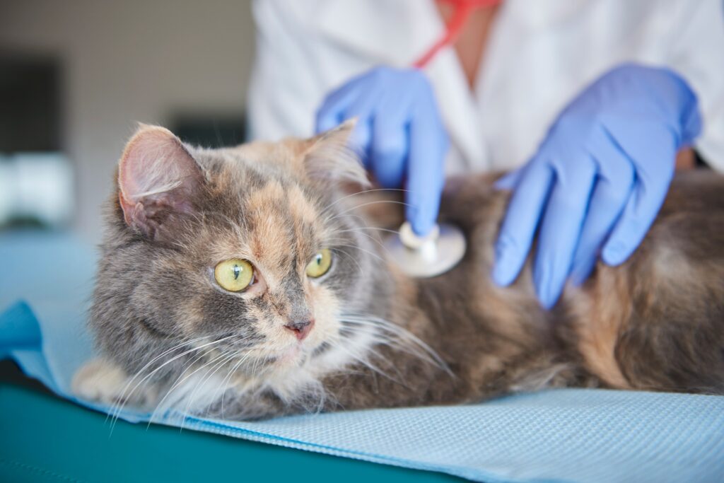
Feline Asthma occurs when the immune system is triggered by allergens causing coughing, wheezing, or difficulty or rapid breathing. The condition is diagnosed by performing bloodwork, fecal examination, heartworm antigen/antibody testing, chest x-rays, cardiac ultrasound, and bronchoscopy with cytology. Treatment may include oxygen therapy, and anti-inflammatory and bronchodilating medications. Chronic therapy with aerosolization of anti-asthmatic medication may be indicated.
Laryngeal Masses can be a benign or malignant proliferation of laryngeal tissue which can lead to acute or chronic upper airway obstruction. Clinical signs may include difficulty breathing, voice change, cough, exercise intolerance, gagging, hypersalivation, collapse, or noting a mass in the neck. The condition is diagnosed by performing bloodwork, neck and chest x-rays, laryngoscopy, ultrasound, or advanced imaging (CT scan or MRI). Treatment includes oxygen therapy, tracheostomy, surgical removal, or radiation therapy.
Laryngeal Paralysis is the lack of opening of the vocal folds and arytenoid cartilages as a result of their associated muscle or nerve dysfunction. Clinical signs include voice change, coughing or gagging when eating, exercise intolerance, difficulty or “raspy” breathing, and collapse. Diagnostic testing may include bloodwork, chest x-rays, thyroid blood testing, examination of the larynx during light sedation, or laryngoscopy. Treatment may involve oxygen therapy, reducing laryngeal edema, and sedation in severe cases. Nonsurgical long term management includes weight loss, restricting exercise, reducing stress, and prevention of hyperthermia in warm conditions. Surgery may be indicated depending on severity. This involves suturing open one or both of the cartilages. There is a lifelong risk of aspiration pneumonia following surgical treatment.
Long Lobe Torsion is the rotation of a lung lobe along its axis usually associated with pleural effusion. Diagnostic testing includes bloodwork, chest x-rays, chest ultrasound, cytology of the pleural fluid, and possibly CT scan. Therapy is directed at stabilizing the patient, improving respiratory function, and removing the affected lung lobe.
Lung Parasites are worms that develop in the major airways or lungs after infection with the larval form of the worm. Clinical signs of infection include coughing, wheezing, exercise intolerance, or respiratory distress. Diagnosis is by fecal testing, Baermann fecal testing, chest x-rays, bloodwork, or bronchoscopy and tracheal or bronchial washes. Treatment includes supportive care and paraciticidal medication.
Mediastinal Disease occurs when air, inflammation, fluid, or a mass develop in the space between the right and left sides of the lungs. Clinical signs can include reluctance to eat, difficulty eating, regurgitation, lethargy, coughing, difficulty breathing, or facial, neck, and/or forelimb swelling. Diagnostic testing involves chest and abdominal x-rays, bloodwork, chest ultrasound, or cytology and culture of the area. Therapy is directed at treating the underlying cause.
Nasal Neoplasia is cancer (usually malignant) occurring in the nasal passages. Clinical signs include hemorrhagic nasal discharge (either one-sided or two-sided), sneezing, reverse sneezing, difficulty or open-mouth breathing, or obvious facial swellings or abnormal appearance to the eye. The condition is diagnosed with bloodwork, lymph node cytology, chest x-rays, nasal imaging (MRI or CT scan), rhinoscopy, or biopsy of affected region. Treatment involves controlling hemorrhage, pain medication, antibiotics for secondary infection, radiation therapy, or chemotherapy.
Nasopharyngeal Polyp is a benign pedunculated mass that originates in the from the middle ear. This occurs primarily in young adult cats. Clinical signs may include nasal discharge, congestion, sneezing, head shaking, pawing at ears, head tilt, malodor to ears, difficulty breathing, or difficulty eating. Diagnostic testing includes an otic exam and cytology, skull x-rays, bloodwork, oral exam under anesthesia, CT scan, and biopsy of the mass. Treatment involves oxygen therapy if respiratory distress is present, treating secondary ear infections, and, ultimately, surgical removal of the mass.
Nasopharyngeal Stenosis is the formation of a thin fibrous membrane at the internal nasal meatus causing narrowing which leads to upper respiratory tract disease. Clinical signs include nasal discharge, sneezing, and worsening of respiratory signs when eating or swallowing. Diagnosis is based on identifying the fibrous membrane with CT scan or pharyngeal exam with bronchoscopy or a dental mirror. Therapy includes removal or dilation of the membrane and anti-inflammatory medication.
Pleural Effusion is the accumulation of fluid in the pleural space in the chest. This can occur as a result of heart disease, infection, inflammation, trauma, protein loss, or cancer. Clinical signs range based on severity of the fluid build up. They can include cough, lethargy, difficulty or rapid breathing, weight loss, anorexia, or distended abdomen. The condition is diagnosed by performing bloodwork, chest and abdominal x-rays, cytology and/or culture of the fluid, or chest ultrasound. Treatment is targeted at supportive care, removal of the fluid, and treating the underlying cause.
Pneumonia, Aspiration can occur when gastric contents or other materials are inhaled into the lungs. Clinical signs include cough, lethargy, difficulty or rapid breathing, collapse, anorexia, or respiratory distress. Diagnostic testing involves bloodwork, chest x-rays, evaluation of blood oxygenation levels, transtracheal lavage for cytology and culture, or bronchoscopy. Therapy includes supportive care (oxygen therapy and intravenous fluids), bronchodilating and antimicrobial medications, courage, and/or nebulization.
Pneumonia, bacterial occurs when there is inflammation of the lungs as a result of a bacterial infection. Clinical signs include cough, nasal discharge, sneezing, lethargy, difficulty or rapid breathing, collapse, anorexia, or respiratory distress. Diagnostic testing involves bloodwork, chest x-rays, evaluation of blood oxygenation levels, transtracheal lavage for cytology and culture, or bronchoscopy. Therapy includes supportive care (oxygen therapy and intravenous fluids), bronchodilating and antimicrobial medications, courage, and/or nebulization.
Pulmonary Edema (Noncardiogenic) occurs when fluid from the vessels builds up in interstitial and alveolar space of the lungs as a result of upper airway obstruction, electrocution, trauma, sepsis, non-septic inflammatory disease, smoke inhalation, or near-drowning experience. Clinical signs include cough, difficulty or rapid breathing, open mouth breathing in cats, weakness, lethargy, decreased appetite, and collapse. The condition is diagnosed with a known underlying cause, a lack of auscultation of a heart murmur, presence of pulmonary crackles or wheezes on exam, chest x-rays, bloodwork, and urine analysis. Therapy is directed at stabilizing the patient with oxygen therapy and bronchodilating medication.
Pulmonary Hypertension (arterial) is elevated arterial pulmonary pressure, which can occur primarily or secondarily to another cause. Underlying causes may include kidney disease, Cushing’s disease, cancer, pancreatitis, heartworm disease, or pulmonary thromboembolism. Clinical signs include cough, difficulty or rapid breathing, abdominal distention, weakness, lethargy, decreased appetite, and collapse. The condition is diagnosed by performing bloodwork, heartworm testing, chest and abdominal x-rays, fecal and Baermann testing, coagulation profile, and chest ultrasound. Therapy includes supportive care and treating the underlying cause for the condition.
Pulmonary Eosinophilic Infiltrates occurs when a particular type of white blood cell (eosinophil) infiltrates the respiratory tract in high numbers without a known underlying cause. Clinical signs include a chronic cough, nasal discharge, exercise intolerance, lethargy, and decreased appetite. Diagnostic testing involves bloodwork, urine analysis, fecal and Baermann testing, chest x-rays, bronchoscopy with cytology and culture, and heartworm testing. Treatment can include steroid medication or removal of solitary mass-forming infiltrates.
Pulmonary Lymphoid Granulomatosis occurs as a rare cancer of infiltrates of lymphoid cells in the lungs. Clinical signs include a cough, exercise intolerance, lethargy, decreased appetite, fever, abdominal distention, enlarged peripheral lymph nodes, or vomiting. Diagnostic testing involves bloodwork, urine analysis, fecal and Baermann testing, chest x-rays, bronchoscopy with cytology and culture, and heartworm testing. Treatment can includes supportive care and chemotherapy.
Respiratory Foreign Body occurs when foreign material lodges in the airways causing obstruction and inflammation. Clinical signs are dependent on location. Nasal foreign body: Sneezing, pawing at face, nasal discharge from one nares, difficulty breathing, or malodor to breath. Tracheal or bronchial foreign body: cough, difficulty breathing, malodor to breath, coughing blood, retching or vomiting, exercise intolerance, or respiratory distress. Bronchiole or pulmonary foreign body: cough, difficulty or rapid breathing, exercise intolerance, lethargy, anorexia, fever, and weight loss. Diagnostic testing includes bloodwork, skull, neck, or chest x-rays, advanced imaging, or bronchoscopy (with cytology and culture). Treatment is directed at supportive care, removing the foreign material, and treating secondary inflammation and infection.
Reverse Sneezing is a noisy paroxysmal sneeze that occurs in the nasopharyngeal region. The condition occurs as a result of an irritant from the environment, nasal mites, infection, or cancer. Diagnostic testing may include bloodwork, viral blood testing, skull x-rays, nasal flush with cytology and culture, rhinoscopy, or CT scan/MRI. Therapy involves treating the underlying cause: anti-inflammatory, antihistamine, or antimicrobial medication, treatment for mites, or treatment for cancer.
Rhinitis, Bacterial is the bacterial inflammation of the one of both of the nasal cavities as a result of another cause. Clinical signs that may be seen include sneezing, nasal discharge, nose bleed, pawing at the nose, head shaking, decreased appetite, or bad breath. Diagnosis is made with bloodwork, oral exam, dental and/or skull x-rays, nasal flush or rhinoscopy with cytology and culture, biopsy, or advanced imaging. Therapy is directed at treating the underlying cause, mucolytic medication, antimicrobial medication, and clearance of nasal secretions.
Rhinitis, Lymphoplasmacytic is a gradually progressive inflammatory nasal disease as a result of infiltration with lymphocytes and plasma cells. The cause is unknown. Clinical signs include sneezing, nasal discharge, nose bleed, pawing at the nose, head shaking, decreased appetite, or ocular discharge. Diagnostic testing includes bloodwork, viral blood testing, imaging of the nasal cavity and sinuses (x-ray, CT scan, or MRI), rhinoscopy with cytology and culture, or biopsy. Treatment includes mucolytic medication, low-dose steroid medication, and antimicrobial medication for secondary bacterial infections.

Sinusitis/Sinus Disorders is mucosal inflammation of one or more of the sinuses as a result of infection, trauma, dental abscess, cancer, or unknown cause. Clinical signs include nasal discharge, sneezing, facial swelling or deformity, lethargy, sensitivity to head, and anorexia. Diagnostic testing often includes bloodwork, viral blood testing, skull x-rays, advanced imaging of skull (CT scan or MRI), rhinoscopy or trephination for cytology, culture, and/or biopsy. Therapy is directed at supportive care and treating the underlying cause.
Tracheal Avulsion is a disruption or tear in the trachea. Clinical signs include subcutaneous emphysema, difficulty or rapid breathing, or respiratory distress. The condition is diagnosed based on recent known trauma, physical exam, neck and chest x-rays, or tracheoscopy. Therapy involves supportive care, treating the underlying cause, or surgical repair of the defect.